
Want to learn more about our solutions?
Contact the VUNO team anytime.
Contact the VUNO team anytime.
Presents key findings essential for diagnosing retinal diseases based on fundus images, helping early diagnosis of major eye diseases.
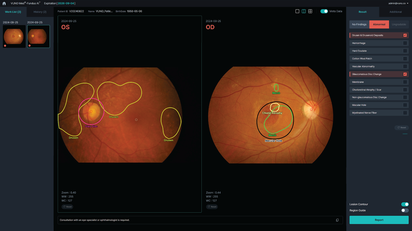
When a fundus photograph is uploaded, the deep learning model analyzes it and identifies 12 abnormal findings, along with their locations, associated with retinal diseases such as diabetic retinopathy, glaucoma, and age-related macular degeneration.
Achieved AUROC values of 96.2%–99.9% across all 12 findings.
*103,262 fundus images with 57 ophthalmologists. Based on internal dataset.
Son J, et al. Development and validation of deep learning models for screening multiple abnormal findings in retinal fundus images. Ophthalmology. 2020;127:85-94. https://doi.org/10.1016/j.ophtha.2019.05.029
Reading time was reduced for both specialists (2%) and residents (26%).
Son J, et al. Development and validation of deep learning models for screening multiple abnormal findings in retinal fundus images. Ophthalmology. 2020;127:85-94. https://doi.org/10.1016/j.ophtha.2019.05.029
AI-assisted reading improved sensitivity by 10% to over 30% compared to unassisted reading, with significant increases across all 12 findings.
Shin JY, et al. Clinical utility of deep learning assistance for detecting various abnormal findings in color retinal fundus images: a reader study. Translational Vision Science & Technology. 2024;13(10):34. https://doi.org/10.1167/tvst.13.10.34

Links to news about VUNO (excluded from medical device advertisement review)
This section is excluded from medical device advertisement review; it links to news related to our company.
This product is designed with high sensitivity to identify potential lesions. The sensitivity can be adjusted according to the usage environment and reading criteria.
This product is an AI-based diagnostic-assist software that supports medical professionals' diagnosis. Final diagnosis and interpretation must be made under the judgment of medical professionals, and it is recommended to use it for screening and reading assistance purposes.
This product is a Class 3 medical device approved by the Ministry of Food and Drug Safety (MFDS), and result reports can be utilized within the approved intended use and scope. However, final interpretation and diagnosis must be based on the judgment of medical professionals, and we recommend applying actual operational procedures according to the hospital's internal processes.
De-identified data was used during the development phase, and personal information was not used in the training process. Clinical data is used solely for diagnostic purposes, and patient data is not used for additional training.
Ad review no.: 62026-|10-31-3900 (valid until 2029-09-15)│This product is a ‘medical device’. Read the ‘precautions for use’ and the ‘instructions for use’ carefully before use.│Category : Digital medical device│Intended use : Software that automatically analyzes abnormal findings in fundus images, presents normal/abnormal results and, when abnormal, indicates the finding and its location to assist clinicians' diagnosis. The product automatically analyzes 12 findings: hemorrhage, cotton wool patch, chorioretinal atrophy, drusen, hard exudate, macular hole, myelinated nerve fiber, membrane, retinal nerve fiber layer defect, glaucomatous disc change, vascular abnormality, and non-glaucomatous disc change